Hip Labral Repair
The labrum of the hip is a fibrocartilaginous structure that attaches to the rim of the acetabulum (socket) providing shock absorption, force distribution, and stability among other functions.
Most hip labral tears occur as a result of underlying femoroacetabular impingement (FAI), commonly referred to as hip impingement, although a subset of athletes can have traumatic labral tears due to one particular event.

Presentation
Most patients will present with a progressive ache in the front and/or side of the hip or groin region. A subset can experience more buttock pain but this is less common. The pain is typically exacerbated with activity, particularly those involving hip flexion. Many patients experience pain with simply sitting for prolonged periods of time.
Work Up
During the initial evaluation of the hip, in addition to the physical examination, X-rays are typically obtained to evaluate the structure of the hip and assess for underlying femoroacetabular impingement or FAI.
Presentation
Nonsurgical
Treatment initially consists of non-operative management with activity modification, NSAIDs, and physical therapy. Non-operative management is typically trialed for 3-6 months depending on symptoms and severity of the deformity. The patients with more severe impingement or deformities tend not to do as well with non-operative management as the deformity is driving the entire process. However we trial everyone with nonsurgical treatment before making a decision for surgery.
Surgical
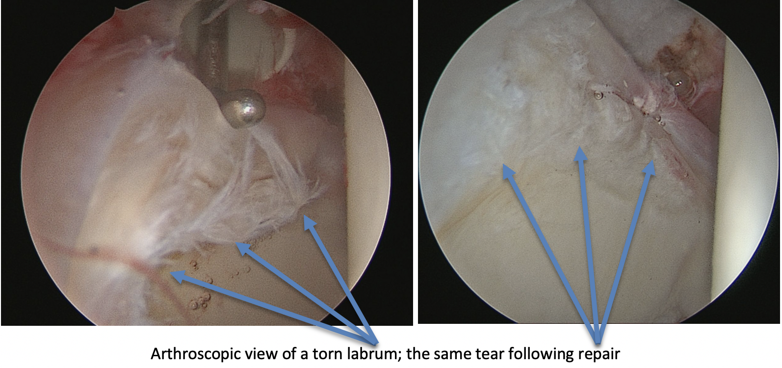
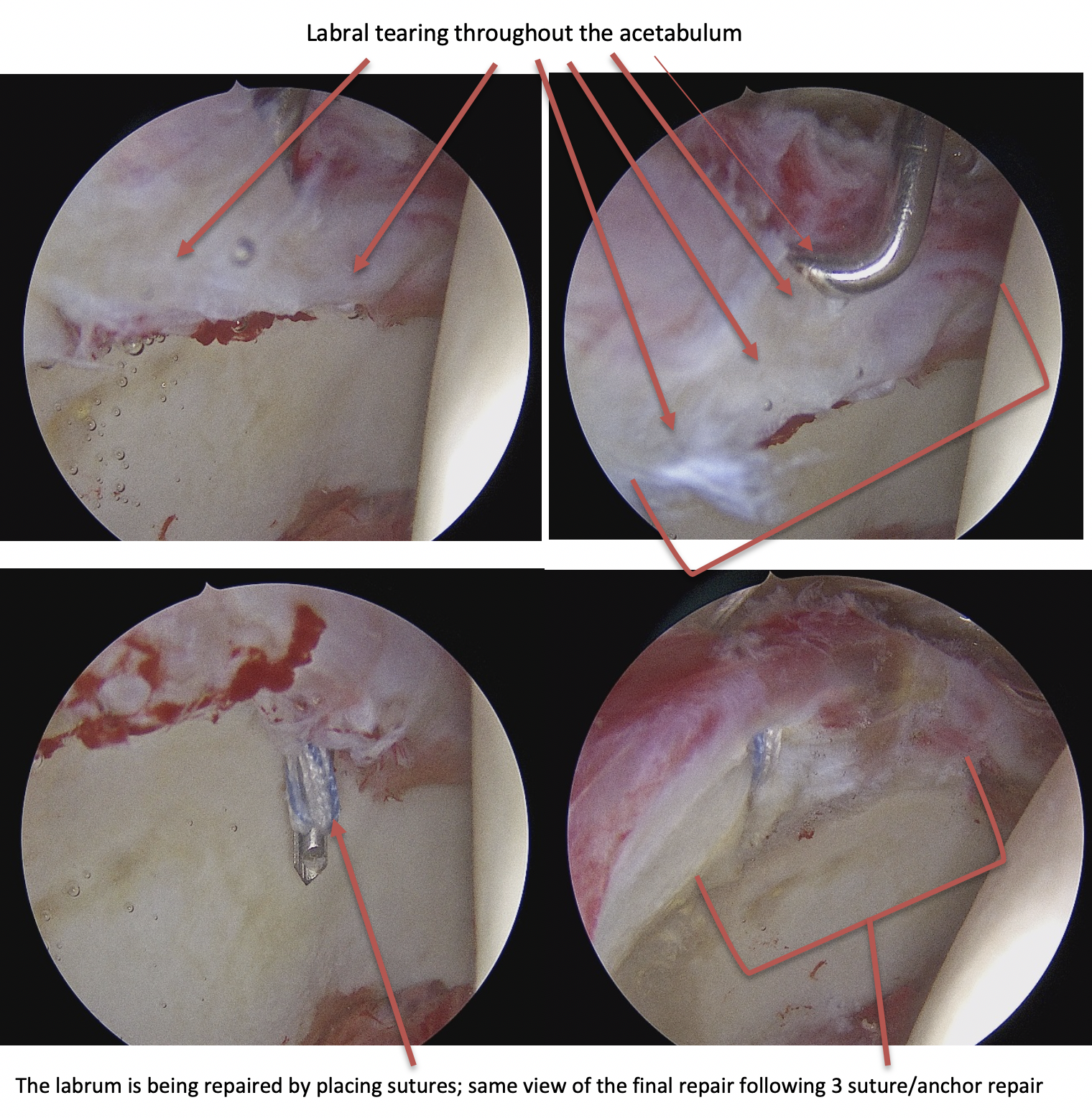
Surgical treatment of labral tears is performed arthroscopically with small incisions. We use an arthroscope (camera) and different instrumentation and anchors to sew the labrum back to the acetabular rim. Any bony deformity (FAI) is addressed at the same time to reshape the hip to a normal shape, thereby protecting the labrum moving forward with the idea of preserving the joint and protecting against arthritis or joint break down.

Recovery
Recovery involves physical therapy beginning a few days after surgery for the first several months in order to regain motion, function and minimize scar tissue formation. Crutches in a partial weight bearing fashion are utilized for the first 2 weeks with progression to weight bearing as tolerated after the 2-week period. Most patients are walking normally 3-4 weeks after surgery. A stationary bike can be started a few days after surgery, elliptical machine typically around 6 weeks, and running progression around 12 weeks. Full recovery and return to sport is typically about 5-6 months following the procedure.
Videos: